2024 - SEMPA reference sample with 3D magnetization
Relevant to TAČR NCK2 project TN02000020/009 within Center of electron and photonic optics
Output n° TN02000020/009-V02, Testing device for out-of-plane (3D) magnetization imaging
Sample contains CoNiB magnetic nanotubes lying on Si (5x5 mm2) substrate (Fig. 1). The tubes have diameters 200-300 nm and host 3D azimuthal magnetization (curling, “vortex-like” domains). These magnetic textures have a well-defined structure with in-plane magnetization on the top surface and opposite out-of-plane magnetization at edges (Fig. 2). The curling sense alternates in neighbouring magnetic domains. Even after saturation in external magnetic field, at remanence, these curling domains are re-established.

Fig. 1 : CoNiB nanotubes dispersed on a Si substrate. Left: Bundle of nanotubes with tube openings. Right: Isolated nanotube with 200nm diameter suitable for magnetic imaging (see Fig. 3).

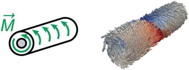
Fig. 2: Azimuthal (curling, “vortex-like”) magnetization in nanotube. Left: Scheme. Right: Visualization of micromagnetic modelling with two domains with opposite curling sense.
The nanotubes were prepared by electroless plating in nanoporous polycarbonate membrane which define their geometry (outer diameter, maximum length). The polymer matrix was then dissolved in organic solvent (dichloromethane). After purification and isolation of nanotubes, they were dispersed on a Si support substrate.
The sample is intended for testing and calibration (alignment) of Scanning Electron Microscopy with Polarization Analysis (SEMPA / spin-SEM). This is originally ultra-high-vacuum-based microscopy technique featuring a detector that measures spins of electrons emitted from magnetic surfaces. SEMPA measures projections of magnetization with very high resolution - down to 10 nm (3 nm with special instrumentation).
Our sample with well-defined 3D magnetization provides both in-plane and out-of-plane magnetization in different sample parts (top surface vs edges; see Fig. 3). SEMPA is sensitive to two orthogonal projections – typically two in-plane directions. However, by tilting the sample, we can access also the out-of-plane component and/or vary relative sensitivity to in-plane and out-of-plane components. In conjunction with micromagnetic simulations, we can obtain even quantitative information regarding magnetization projection onto the detector axes.
The special magnetic configuration of curling domains enables to detect uniquely out-of-plane magnetization component in one channel, whereas mixture of in-plane and out-of-plane components is typically detected by SEMPA unless so called spin-rotator is available (additional costly extension of the detector, often not present).

Fig. 3 : SEMPA magnetic imaging of a CoNiB nanotube on a Si substrate (sample tilt 45 deg). Topography (normal electron micrograph) and two magnetic projections are shown. The first would be mixture of longitudinal Mx (in-plane, along tube) and out-of-plane Mz components. But thanks to the special magnetic texture, there is no Mx in domains (confirmed by magnetic force microscopy and synchrotron imaging with polarized X-Rays) and only Mz projection is imaged (weak, opposite contrast at edges; alternating in each domain). The second projection is transverse with maximum near the tube top surface and zero at edges.


